Welcome to the Sakellariou Laboratory on Magnetic Resonance
We are a research group on nuclear magnetic resonance spectroscopy, relaxometry, and imaging. Our background comes from solid-state NMR and low-field instrumentation.
Our mission is to develop innovative magnetic resonance approaches for real-world applications in various domains, including sustainable chemistry and material science, geology, and biomedicine.
A brief introduction
The Sakellariou Laboratory of Magnetic Resonance is part of KU Leuven in Leuven, Belgium, in the Faculty of Bioscience Engineering and of the Centre for Membrane Separations, Adsorption, Catalysis and Spectroscopy for Sustainable Solutions of Surface Science and Catalysis (cMACS). We are also members of the KU Leuven Institutes for Sustainable Metals and Minerals (SIM2) and Micro- and Nanoscale Integration (LIMNI).
We used to be part of the Laboratory of Structure and Dynamics by Magnetic Resonance, which is part of the Saclay Institute of Matter and Radiation and belongs to the Direction of Fundamental Research of the French Atomic Energy Commission (C.E.A.) (Saclay, France).
Our research themes
Our research themes span various magnetic resonance techniques in both low-field and high-field NMR, as detailed below. For further inquiries or interest in collaborations, please contact us.
Novel imaging technologies and methodologies:
Magnetic resonance imaging in a unique non-invasive technique to “see” inside opaque three dimensional objects. Our lab has been developing novel low- and ultra-low field magnetic resonance imaging sensors for small animals in the framework of the GammaMRI project for enhancing the signal sensitivity using photon-detected magnetic resonance and hyperpolarization. Furthermore, we have been working to improve magnetic field quality and to produce specifically shaped magnetic fields using 3D-printed shim correctors for preclinical and clinical MRI within the 3DPBrain project. Spectral resolution in MRI often suffers from anisotropies and we have been developing a unique spinning magnet technology in the frame of the ERC project R-EvolutioN-M-R to solve these issues and record ultra-high-resolution chemical shift resolved imaging in vivo.
In situ MR monitoring of chemical processes:
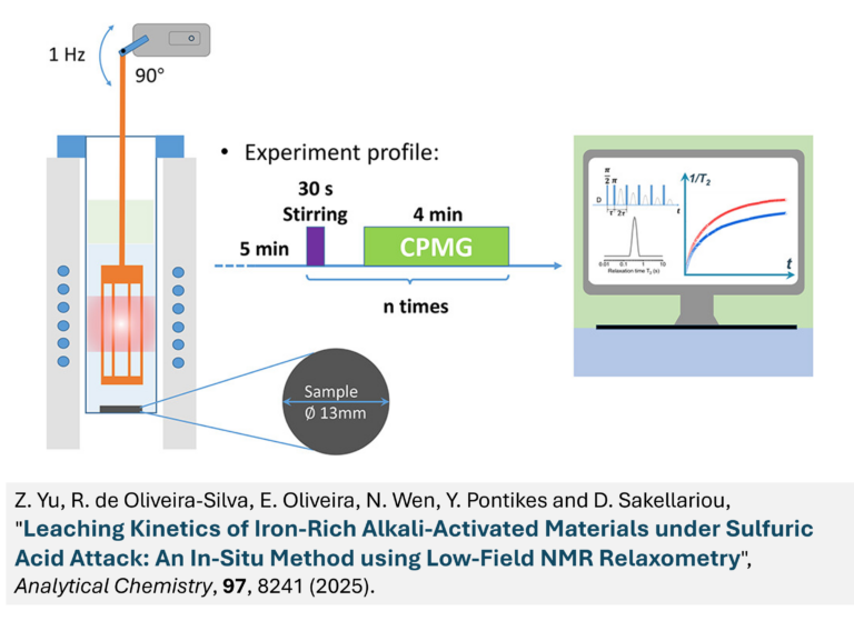
Modern high-field magnetic resonance cannot be easily adapted for in situ, operando applications relevant for industrial applications. We develop low-field magnetic resonance sensors that measure relaxation and diffusion in samples that may have specific spatial, temperature, pressure, etc. requirements. For such systems, we build the Magnetic resonance sensor around the sample or device to be studied. One such application for geopolymer monitoring during curing was developed within the PorMedNMR project.
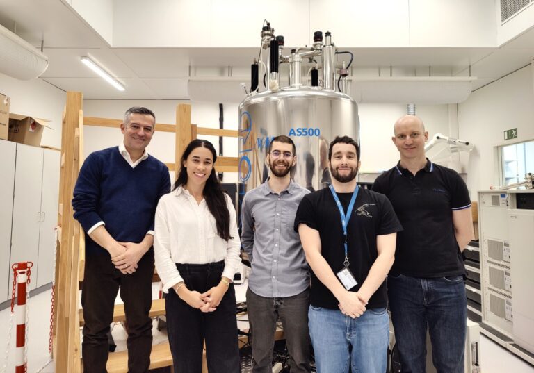
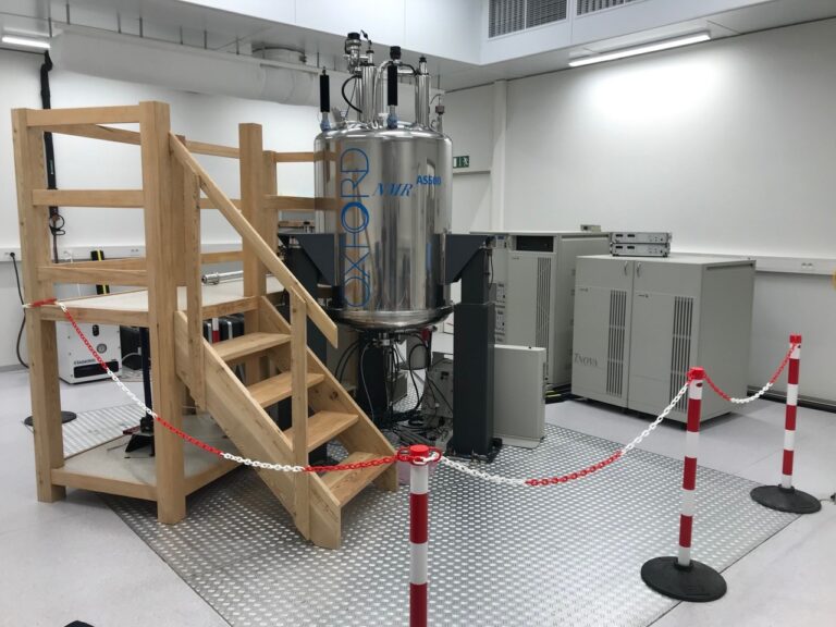
Material characterisation with high-field NMR spectroscopy (in situ and ex situ):
Solid-state NMR techniques provide unique information on the structure and molecular dynamics of non-isotropic and heterogeneous samples, even when long-range order is absent (amorphous). For this reason, they are an indispensable tool for materials characterisation, covering a wide range of applications, from organic and inorganic powdered solids, energy, construction, and geological materials, to biominerals, tissues and cells, polymers, and food products.
A notable intercommunity project we have had the chance to participate in (Tethered) focuses on the characterization of soft polymers having hidden lengths and self-healing properties. Within this project, a variety of characterization approaches (including high- and low-field NMR) are employed to shed light on these complex materials.
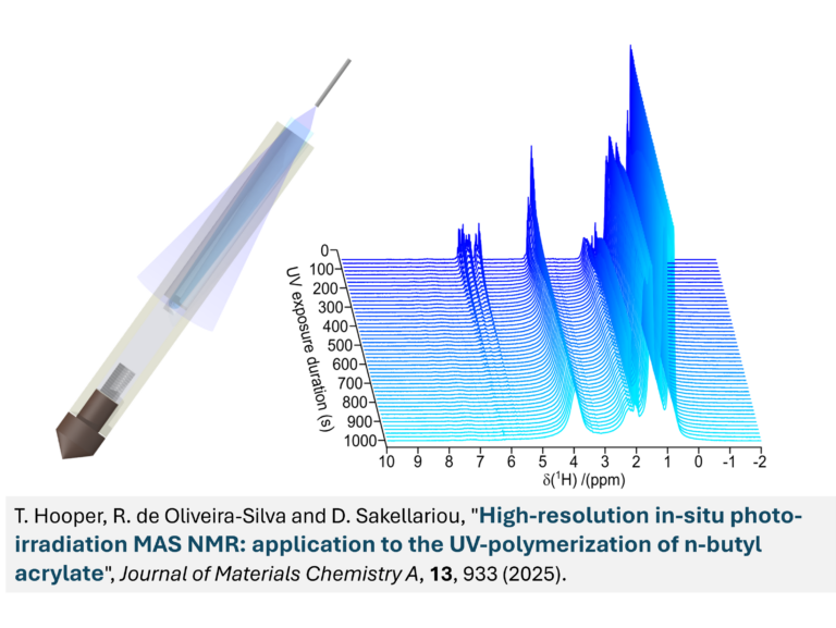
We are also optimising methodologies for in situ solid-state NMR characterisation of light-activated materials within the FWO postdoctoral project 1253824N. We have been able to monitor the chemical reaction of photo-polymerization initiated with laser irradiation under Magic Angle (sample) Spinning (MAS). This approach can be applied in light-irradiated materials with particular focus on solar cells and perovskites.
Molecular sorption monitored in situ by Magnetic Resonance:
Sorption in porous media is essential for tailoring the physical and chemical properties of materials in many applications, such as separations, catalysis, etc. Metal-organic frameworks (MOFs), zeolites, food products, and many other materials exhibiting micro- and meso-porosity are usually studied using gas or vapour sorption volumetric analysis. We have developed an in situ approach, named MR-Relaxorption, to study sorption in which relaxation is measured in operando throughout the complete sorption/desorption isotherm.
Programmable and magnetically controlled biological materials:
Can magnetic fields be used to actuate, shape, and fabricate living organisms? In this multidisciplinary field of research, we aim to shape in space and control time-dependent magnetic fields. This work is within the frame of collaborative projects Organdroids and CartiBot.
Micro-detection with Magic Angle Coil Spinning:
Magnetic Resonance at room temperature intrinsically has a rather low signal-to-noise ratio. When studying microscopic samples, this renders the data acquisition lengthy or even impossible. To improve the detection limits in static and spinning samples of nanoliter volumes, we work on the development of Magic Angle Coil Spinning (MACS) techniques.
Portable Magnetic Resonance:
There are many cases where the sample or object of study cannot be analysed using standard NMR and MRI hardware due to space constraints. Low-field NMR offers unique advantages in terms of applicability outside the laboratory and cost. It has been the playground for original instrumentation in our laboratory since 2007, when the NMR2GO project was funded to develop portable NMR sensors.
Today, we have a variety of home-made benchtop systems offering unique and modular possibilities for analysing samples that cannot be measured with commercial NMR hardware.
Ultra-slow Magic Angle Spinning:
Magic Angle (sample) Spinning (MAS) has been the cornerstone of solid-state NMR because it yields isotropic, highly resolved peaks when the sample is spun rapidly. There are cases, however, where such fast spinning is unwanted, for example, when the sample is sensitive, large, and/or alive! In such cases, slow and ultra-slow spinning techniques should be used, while ensuring that spinning sidebands are eliminated and isotropic peak information is obtained.
Our funding
We are thankful for the generous funding received from the following funding agencies: